Anatomy Of Chest - In insects, crustaceans, and the extinct trilobites, the thorax is one of the three main divisions of the creature's body, each of which is in turn composed of multiple segments.
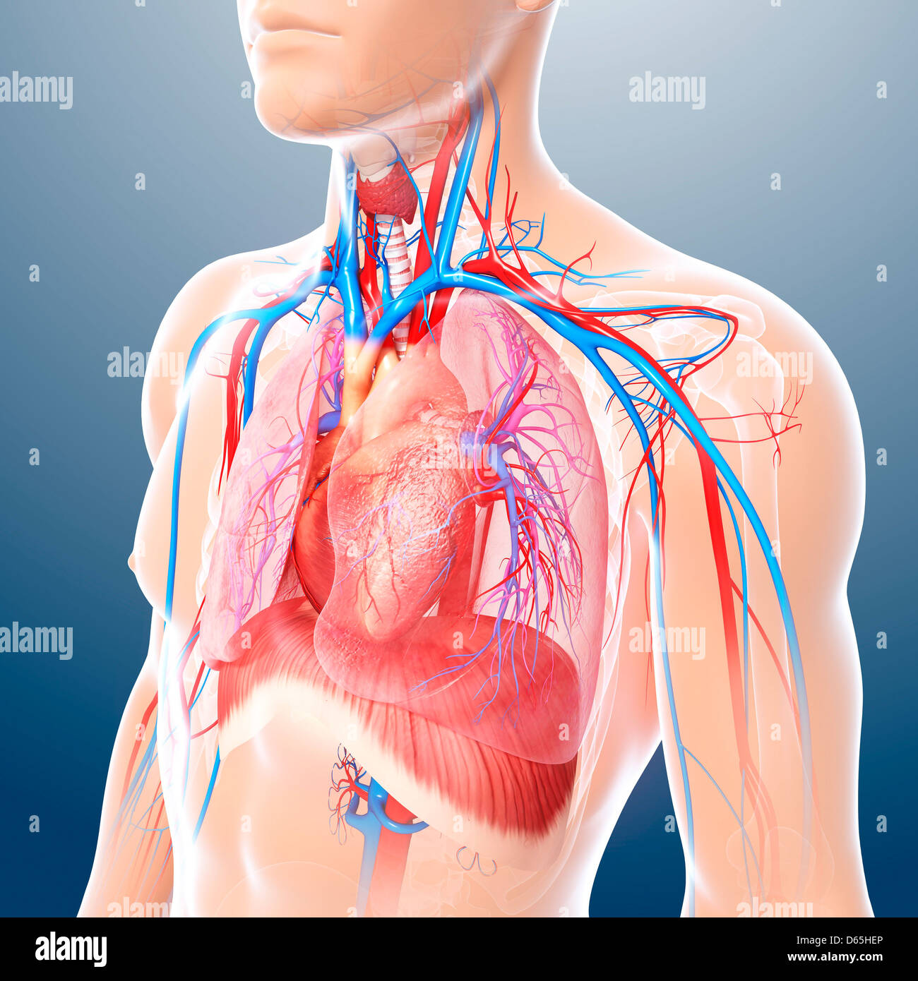
Anatomy Of Chest - In insects, crustaceans, and the extinct trilobites, the thorax is one of the three main divisions of the creature's body, each of which is in turn composed of multiple segments.. The anatomic illustrations are presented as… About the 6th week, the somites differentiate into the sclerotomes and the dermatomyotomes. 2 skin of the anterior chest wall syllabus p. The circulatory system does most of its work. The chest is the area of origin for many of the body's systems as it houses organs such as the heart, esophagus, trachea, lungs, and thoracic diaphragm.
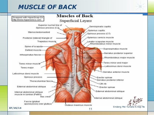
The chest anatomy includes the pectoralis major, pectoralis minor and the serratus anterior. The chest is made up primarily of two muscles: Anatomy of the chest, abdomen, and pelvis was produced in part due to the generous funding of the david f. You will also find the xiphoid process, 10th rib, the apex of the heart, the coronary vein of the heart. Here, we break down the anatomy of your chest muscles.
M Of Chest Abd Back Semester 2 Kd 2 Anatomy from image.slidesharecdn.com See chest anatomy stock video clips. Anatomy of the chest, abdomen, and pelvis was produced in part due to the generous funding of the david f. The chest wall is comprised of skin, fat, muscles, and the thoracic skeleton. Abdominal regions and organs 12 photos of the abdominal regions and organs 9 abdominal regions and its organs, abdominal cavity regions and organs, abdominal regions and associated organs, abdominal regions and its organs, abdominal regions and quadrants and organs, human anatomy, 9 abdominal regions and its organs. Applied anatomy of the chest wall and mediastinum petros mirilas michael e. This chapter is an abbreviated review of thoracic anatomy as seen on chest radiographs and computed tomography (ct) of the chest. The chest or thorax is the region between the neck and diaphragm that encloses organs, such as the heart, lungs, esophagus, trachea, and thoracic diaphragm. Here, we break down the anatomy of your chest muscles.
Here, we break down the anatomy of your chest muscles.
Basic thoracic anatomy and physiology an understanding of thoracic imaging requires knowledge of the anatomy being imaged, as described in this chapter, as well as the imaging techniques applied to the thorax, covered in chapter 2. Muscles of the chest and their functions you have two mighty muscles on both sides of your chest: 31 anatomy of the female breast syllabus p. Radiological anatomy of the lungs, mediastinal lymph nodes, trachea, bronchi, pleural cavity, heart and pulmonary vessels. The epidermis is the outermost layer that provides a protective, waterproof seal over the body. Anatomy of the thorax, heart, abdomen and pelvis recommended text gray's anatomy for students. Anatomy of pancreas 12 photos of the anatomy of pancreas anatomy and histology of pancreas ppt, anatomy and physiology of human pancreas, anatomy of pancreas divisum, describe the microscopic anatomy of pancreas, gross anatomy of pancreas, human anatomy, anatomy and histology of pancreas ppt, anatomy and physiology of. Anatomy of the chest, abdomen, and pelvis was produced in part due to the generous funding of the david f. Computed tomography (ct) of the chest can detect pathology that may not show up on a conventional chest radiograph(1). Three dimensional view of the female reproductive system, full frontal view. 12 cm (5 in) in length, 8 cm (3.5 in) wide, and 6 cm (2.5 in) in thickness. Here, we break down the anatomy of your chest muscles. The anatomic illustrations are presented as…
See chest anatomy stock video clips. Anatomy of the thorax, heart, abdomen and pelvis recommended text gray's anatomy for students. Browse 6,407 chest anatomy stock photos and images available, or search for human anatomy to find more great stock photos and pictures. Basic thoracic anatomy and physiology an understanding of thoracic imaging requires knowledge of the anatomy being imaged, as described in this chapter, as well as the imaging techniques applied to the thorax, covered in chapter 2. Plus, how to target each to make them bigger and stronger.
Chest Anatomy Artwork Stock Photo Alamy from c8.alamy.com Hemi diaphragm normal chest anatomy lateral chest xray colon gas trachea oblique fissure horizontal fissure rt. Normal anatomy of the thorax on labeled chest ct: Three dimensional view of the female reproductive system, full frontal view. The chest anatomy includes the pectoralis major, pectoralis minor and the serratus anterior. See chest anatomy stock video clips. Plus, how to target each to make them bigger and stronger. See human chest anatomy stock video clips. 4 innervation of the breast blood supply of the breast syllabus p.
The circulatory system does most of its work.
Radiology basics of chest ct anatomy with annotated coronal images and scrollable axial images to help medical students and junior doctors learning anatomy. Learn about each of these muscles, their locations, functional anatomy and exercises for them. The pectoralis major and the pectoralis minor, known collectively as your pecs. Download my two educational text books for free using this link: The circulatory system does most of its work. The first step in understanding thorax anatomy is to find out its boundaries. 4 innervation of the breast blood supply of the breast syllabus p. Chest bone, ribs, lung, heart, xiphoid process, sternum anatomy. See human chest anatomy stock video clips. Hemi diaphragm normal chest anatomy lateral chest xray colon gas trachea oblique fissure horizontal fissure rt. See chest anatomy stock video clips. System respiratory respiratory organs of human body digestive and respiratory system medical chest internal structure of human body medicine body lungs biology intestines stomach anatomy torso human internal. Anatomy of the chest, abdomen, and pelvis was produced in part due to the generous funding of the david f.
Abdominal regions and organs 12 photos of the abdominal regions and organs 9 abdominal regions and its organs, abdominal cavity regions and organs, abdominal regions and associated organs, abdominal regions and its organs, abdominal regions and quadrants and organs, human anatomy, 9 abdominal regions and its organs. Radiology basics of chest ct anatomy with annotated coronal images and scrollable axial images to help medical students and junior doctors learning anatomy. Anatomy of the thorax, heart, abdomen and pelvis recommended text gray's anatomy for students. Normal anatomy of the thorax on labeled chest ct: 12 cm (5 in) in length, 8 cm (3.5 in) wide, and 6 cm (2.5 in) in thickness.
Surgical Anatomy Of The Chest Wall Thoracic Key from thoracickey.com About the 6th week, the somites differentiate into the sclerotomes and the dermatomyotomes. Thoracic cavity, also called chest cavity, the second largest hollow space of the body. Browse 6,407 chest anatomy stock photos and images available, or search for human anatomy to find more great stock photos and pictures. Normal anatomy of the thorax on labeled chest ct: The thorax or chest is a part of the anatomy of humans, mammals, other tetrapod animals located between the neck and the abdomen. Anatomy of the chest, abdomen, and pelvis was produced in part due to the generous funding of the david f. It provides protection to vital organs (eg, heart and major vessels, lungs, liver) and provides stability for movement. The circulatory system does most of its work.
Table 1.1 lists the major anatomic structures within the thorax that are discussed.
Sternocleidomastoid muscle clavicle and ribs anatomy muscle anatomy chest sternocleidomastoid ribs anatomy chest muscles anatomy thorax rib muscles chest muscles chest anatomy illustration. A good radiologist knows the anatomy because knowing where structures normally live and recognizing the location of an abnormality helps to make or narrow the differential diagnosis. 4 innervation of the breast blood supply of the breast syllabus p. Learn about each of these muscles, their locations, functional anatomy and exercises for them. Anatomy of the thorax, heart, abdomen and pelvis recommended text gray's anatomy for students. The circulatory system does most of its work. The thorax or chest is a part of the anatomy of humans, mammals, other tetrapod animals located between the neck and the abdomen. The chest is made up primarily of two muscles: Plus, how to target each to make them bigger and stronger. (1) the pectoralis major, and (2) the pectoralis minor. The chest anatomy includes the pectoralis major, pectoralis minor and the serratus anterior. Swensen fund for innovation in teaching. Three dimensional view of the female reproductive system, full frontal view.


0 Komentar